Fetal Growth Calculator
Track your baby's development • 2026 edition
Fetal Growth Formula:
Show the calculator\( \text{Estimated Weight (g)} = 1.07 \times BPD^3 + 0.3 \times AC^2 \times FL \)
Where:
- \( BPD \) = Biparietal Diameter (cm)
- \( AC \) = Abdominal Circumference (cm)
- \( FL \) = Femur Length (cm)
Alternatively, to calculate gestational age from measurements:
\( \text{Gestational Age (weeks)} = \frac{\text{BPD (mm)} + 10}{10} \)
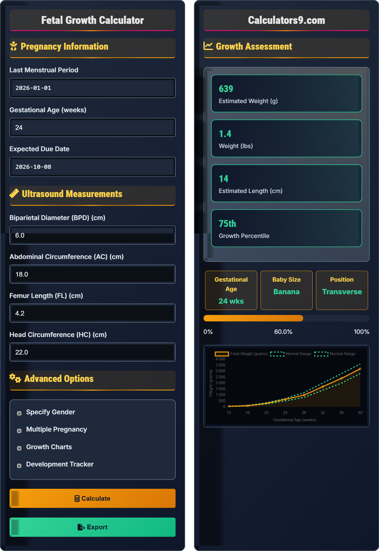
This formula estimates fetal weight using ultrasound measurements. The Hadlock formula (shown above) is widely used by healthcare professionals to assess fetal growth and development. These measurements help monitor baby's growth and identify potential concerns.
Example: If BPD=8.2cm, AC=26.5cm, FL=6.1cm:
Weight = 1.07 × (8.2)³ + 0.3 × (26.5)² × 6.1
= 1.07 × 551.4 + 0.3 × 702.25 × 6.1
= 589.9 + 1285.1 = 1875g (about 4.1 lbs)
This provides an estimated fetal weight at the time of ultrasound.
Pregnancy Information
Ultrasound Measurements
Advanced Options
Growth Assessment
| Measurement | Value | Expected | Percentile |
|---|
| Milestone | Week | Status | Description |
|---|
Growth References
Fetal Growth Monitoring Guide
Fetal growth is monitored throughout pregnancy using various measurements obtained during ultrasounds. These measurements help assess the baby's development and identify any potential growth concerns.
The Hadlock formula is commonly used to estimate fetal weight:
Where:
- \(EFW\) = Estimated Fetal Weight (grams)
- \(BPD\) = Biparietal Diameter (cm)
- \(AC\) = Abdominal Circumference (cm)
- \(FL\) = Femur Length (cm)
Interpretation of growth measurements:
- 5th-95th percentile: Normal growth range
- Below 5th percentile: Small for gestational age
- Above 95th percentile: Large for gestational age
- 10th-90th percentile: Optimal growth zone
- Attend all appointments: Regular ultrasounds are crucial
- Maintain health: Good nutrition supports growth
- Track measurements: Note trends over time
- Ask questions: Understand your baby's growth pattern
- Follow recommendations: Address any concerns promptly
Growth Monitoring Basics
Monitoring baby's development.
\(EFW = 1.07 \times BPD^3 + 0.3 \times AC^2 \times FL\)
Using ultrasound measurements.
- Measurements taken during ultrasounds
- Percentiles indicate normal ranges
- Trends are more important than single values
Monitoring Strategies
Comparing to population norms.
- Regular ultrasound measurements
- Compare to growth charts
- Identify trends over time
- Consult healthcare provider
- Individual variation is normal
- Genetics affect growth patterns
- Multiple pregnancies differ
- Early measurements less accurate
Fetal Growth Monitoring Learning Quiz
Which ultrasound measurement is the best indicator of fetal weight and nutrition?
The answer is B) Abdominal Circumference (AC). The abdominal circumference is the best indicator of fetal weight and nutritional status because it reflects the size of the liver and the amount of subcutaneous fat, both of which are closely correlated with overall fetal mass and nutritional condition.
The abdominal circumference measurement is particularly important because it correlates most closely with fetal weight. Unlike head measurements which primarily reflect brain development, or bone measurements which reflect skeletal growth, the abdominal circumference reflects the visceral organs and fat stores that indicate the baby's overall nutritional status and growth.
AC: Abdominal Circumference measurement
Fetal Weight: Estimated using multiple measurements
Nutritional Status: Baby's nourishment level
• AC best reflects fetal weight
• Combined measurements improve accuracy
• Trend monitoring is crucial
• AC is most sensitive to growth changes
• Monitor AC trends over time
• Low AC may indicate growth restriction
• Relying on single measurements
• Not tracking trends over time
• Overlooking AC significance
If a baby's estimated weight is at the 25th percentile, what does this mean?
The baby weighs more than 25% of babies of the same gestational age and less than 75% of babies of the same gestational age. This is considered within the normal range, as percentiles between 5th and 95th are generally considered normal. The 25th percentile indicates the baby is growing appropriately but on the smaller side of normal.
Growth percentiles compare a baby's measurements to a reference population of the same gestational age. A 25th percentile means that if you lined up 100 babies of the same gestational age by weight, this baby would rank 25th from the lightest. This is perfectly normal and healthy.
Percentile: Relative ranking in population
Reference Population: Standard growth curves
Normal Range: 5th to 95th percentile
• 5th-95th percentile is normal
• Trends matter more than single values
• Individual variation is expected
• Focus on consistent growth patterns
• Small babies can be perfectly healthy
• Genetics influence size
• Panicking over single low percentile
• Comparing to other pregnancies
• Not considering genetic factors
During an ultrasound at 32 weeks, the measurements were: BPD=8.1cm, AC=26.2cm, FL=5.9cm. Using the Hadlock formula, what is the estimated fetal weight? (Formula: EFW = 1.07×BPD³ + 0.3×AC²×FL)
Step 1: Calculate BPD³
BPD³ = 8.1³ = 8.1 × 8.1 × 8.1 = 531.44 cm³
Step 2: Calculate AC²
AC² = 26.2² = 26.2 × 26.2 = 686.44 cm²
Step 3: Apply Hadlock formula
EFW = 1.07 × 531.44 + 0.3 × 686.44 × 5.9
EFW = 568.64 + 0.3 × 4049.996
EFW = 568.64 + 1215.00
EFW = 1783.64 grams ≈ 1784 grams
The estimated fetal weight is approximately 1784 grams (about 3.9 pounds).
This calculation demonstrates how healthcare providers estimate fetal weight using the Hadlock formula. The formula incorporates multiple measurements to provide a more accurate weight estimation than using any single measurement alone. This helps monitor growth and identify potential concerns.
Hadlock Formula: Standard weight estimation method
EFW: Estimated Fetal Weight
Ultrasound Measurements: Tools for growth assessment
• Use multiple measurements for accuracy
• Formula provides estimate, not exact weight
• Compare to expected values for gestational age
• Measurements may vary slightly between technicians
• Accuracy improves in second and third trimesters
• Use as part of overall growth assessment
• Taking single measurements too seriously
• Not accounting for measurement variability
• Ignoring growth trends
A pregnant woman's baby was measured at the 60th percentile at 20 weeks but dropped to the 15th percentile at 24 weeks. What might this indicate, and what should be the next steps?
A drop from the 60th to 15th percentile represents a significant decrease in growth velocity that warrants attention. This pattern suggests possible intrauterine growth restriction (IUGR), where the baby is not growing at the expected rate.
Next steps should include:
• Repeat ultrasound in 2-3 weeks to confirm the trend
• Assess Doppler flow studies to check placental function
• Evaluate maternal factors (nutrition, hypertension, infections)
• Consider additional testing if growth continues to slow
• Increase monitoring frequency
This significant percentile drop indicates the need for closer surveillance.
Monitoring growth trends over time is more important than single measurements. A drop of more than 20 percentile points between ultrasounds is considered significant and requires further evaluation. Healthcare providers look for consistent growth patterns rather than isolated measurements.
IUGR: Intrauterine Growth Restriction
Growth Velocity: Rate of growth over time
Trend Monitoring: Tracking changes over time
• Trends matter more than single values
• Drop >20 percentiles needs evaluation
• Multiple factors affect growth
• Track measurements over time
• Don't panic over single measurements
• Follow provider's recommendations
• Overreacting to single measurements
• Ignoring concerning trends
• Not following up on recommendations
How does fetal growth typically differ in multiple pregnancies compared to singleton pregnancies?
The answer is B) Multiples are typically smaller than singletons. In multiple pregnancies, babies often grow at a slower rate and are born smaller than singletons due to shared resources and space limitations in the uterus. Twins are often considered large if they reach 5.5-6 pounds each, whereas singletons of the same gestational age might weigh 7-8 pounds.
Multiple pregnancies have different growth expectations because the available space and nutrients must be shared between babies. Healthcare providers use different growth charts for multiples and consider smaller sizes normal if the babies are growing proportionally. The uterus has physical limitations that affect growth in multiple pregnancies.
Singleton: Single baby pregnancy
Multiple: Two or more babies
Resource Sharing: Nutrients divided between babies
• Multiples grow differently than singletons
• Smaller size may be normal for multiples
• Increased monitoring is required
• Use multiples-specific growth charts
• Monitor growth more frequently
• Focus on proportional growth
• Comparing multiples to singleton charts
• Assuming smaller multiples are unhealthy
• Not increasing monitoring frequency
FAQ
Q: How accurate are ultrasound measurements for predicting baby's size?
A: Ultrasound measurements have a margin of error of approximately 10-15% for weight estimation. The Hadlock formula:
\( EFW = 1.07 \times BPD^3 + 0.3 \times AC^2 \times FL \)
Provides the most accurate estimation when multiple measurements are used. Accuracy improves in the second and third trimesters. Factors affecting accuracy include baby's position, amniotic fluid levels, and maternal body habitus.
Q: What does it mean if my baby is measuring small?
A: Babies measuring below the 10th percentile may be classified as small for gestational age (SGA). This could be due to:
• Constitutional smallness (genetic factors)
• Intrauterine growth restriction (IUGR)
• Placental insufficiency
• Maternal factors (hypertension, diabetes, lifestyle)
Further evaluation includes serial measurements, Doppler studies, and assessment of amniotic fluid to differentiate between constitutionally small and growth-restricted babies.